 EMBLLM
Why this matters
Acute vision loss is a time-critical complaint: some causes threaten the eye within hours, and some threaten the patient's life. Eye complaints make up up to about 2% of ED visits, yet many emergency clinicians feel least confident here.
This post is built on a 2026 narrative review by Maher, Guo and Yek in Clinical Ophthalmology, which distills the evaluation into five steps: laterality, pain, targeted history, focused chairside exam with red flags, and act (investigate, escalate, refer). The authors are clear that this is not a new algorithm. It is a practical synthesis of existing principles, sized for the ED, primary care and general ophthalmic settings.
The core message: you do not need a definitive diagnosis at the first visit. You need to recognize red flags and get the right specialist involved fast. Investigations should never delay escalation when red flags are present.
Scope note: the review covers adults with acute loss. Pediatric causes and chronic, progressive loss are outside its scope.
Steps 1–3: the history does most of the sorting
Step 1 — One eye or both?
- Monocular loss localizes in front of the chiasm: media, retina or optic nerve.
- Binocular loss points to the chiasm, retrochiasmal pathways or brain.
- Patients often mistake binocular blur (for example, a homonymous field cut) for loss in one eye. Cover each eye and test separately.
- Treat a homonymous field defect as cerebrovascular disease until proven otherwise. Normal pupils and a normal anterior segment with vision loss should raise suspicion for occipital infarction.
- Transient positive phenomena (scintillations, zig-zag lines) in a younger patient with a normal exam suggest migraine aura.
Step 2 — Painful or painless?
| Pain pattern | Think |
|---|---|
| Severe eye pain, headache, nausea/vomiting, halos | Acute angle-closure glaucoma (AACG) |
| Pain on eye movement | Optic neuritis |
| Surface irritation, photophobia | Keratitis, anterior uveitis |
| Sudden and painless | Retinal artery or vein occlusion, retinal detachment, vitreous hemorrhage, ischemic optic neuropathy |
Step 3 — Targeted history
Tempo. Loss that is maximal within seconds suggests vascular occlusion or trauma. Loss over hours to days suggests inflammatory or infectious causes. Transient loss deserves the same urgency as persistent loss when a vascular cause is possible (amaurosis fugax).
Associated symptoms.
| Symptom | Suggests |
|---|---|
| Flashes, floaters, curtain or shadow | Retinal detachment |
| Halos | AACG |
| Transient dimming | Amaurosis fugax |
| Pain with eye movement | Optic neuritis |
| Photophobia | Keratitis, uveitis |
| Headache, jaw claudication, age over 50 | Giant cell arteritis (GCA) |
| Focal neurologic deficits | Stroke |
Recent ocular surgery or intravitreal injection. Decline in vision with pain, redness or hypopyon, typically within 3–5 days of cataract surgery or an injection, is endophthalmitis until proven otherwise. This needs immediate ophthalmology.
Systemic risk factors.
- Diabetes: diabetic macular edema, vitreous hemorrhage, tractional detachment, CRVO.
- Vascular risk (hypertension, hyperlipidemia, carotid disease, smoking): retinal artery and vein occlusion. Up to two-thirds of CRAO patients have cardiovascular or cerebrovascular disease.
- NAION risk: nocturnal hypotension, diabetes, obstructive sleep apnea.
- Autoimmune disease (RA, GPA, sarcoidosis, HLA-B27 disease): uveitis, scleritis, optic neuropathy.
- Immunosuppression (steroids, chemotherapy, biologics, HIV, recent sepsis): severe infection with muted signs. Lower your threshold to call ophthalmology.
Step 4: the focused chairside exam
A systematic bedside exam, done in the same order every time, catches most red flags before any imaging. Run it front to back.
| Component | How | What to look for |
|---|---|---|
| General inspection | Face, lids, orbit | Vesicular V1 rash with Hutchinson sign (zoster ophthalmicus); lid edema, erythema, proptosis (orbital cellulitis) |
| Visual acuity | Each eye separately, with glasses; pinhole if reduced | Improves with pinhole = refractive or media cause. No improvement = organic pathology. Normal acuity does not exclude a dangerous cause. |
| Pupils | Swinging flashlight | RAPD = optic nerve or severe retinal disease. Fixed mid-dilated pupil = AACG, CN III palsy, pharmacologic or traumatic mydriasis |
| Confrontation fields | Each eye, all quadrants | Altitudinal = ischemic optic neuropathy. Homonymous = stroke or retrochiasmal lesion. Central scotoma = optic neuritis. Bitemporal = chiasmal compression (pituitary adenoma or apoplexy) |
| External and anterior segment | Penlight or slit lamp | Injection pattern, discharge, hypopyon, hyphema, corneal opacity, shallow anterior chamber, keratic precipitates |
| Fluorescein | Cobalt blue light | Dendrites (herpetic keratitis), punctate erosions, focal ulcer. Positive Seidel = open globe |
| Fundus | Direct ophthalmoscopy or photography | Cherry-red spot (CRAO), "blood and thunder" (CRVO), disc swelling (papilledema, optic neuritis, hypertensive emergency), grey elevated retina (RD), vitreous haze (hemorrhage, endophthalmitis) |
| Intraocular pressure | Tonometry | Raised in AACG. Skip it if globe rupture is possible. |
Two exam pearls worth repeating. A patient complaining of "vision loss" with 20/20 acuity may have a field defect, so always check fields. And a macula-on retinal detachment can cause peripheral field loss while central acuity is still preserved, which is exactly the window when surgery saves the most vision.
When you cannot see the fundus. B-scan ultrasound helps when the view is obscured, identifying retinal detachment, vitreous pathology and the T-sign of posterior scleritis. (The review does not discuss emergency-physician ocular POCUS specifically; many EDs use it for the same questions, and findings should follow your local credentialing and protocols.)
Step 5: act on the can't-miss diagnoses
Each of these has a recognizable pattern and a defined first move in the ED. Escalation comes first; the workup follows or runs in parallel.
| Diagnosis | Key features | ED action |
|---|---|---|
| Central retinal artery occlusion (CRAO) | Sudden, profound, painless monocular loss; RAPD; cherry-red spot | Activate the stroke pathway (CRAO is a stroke equivalent); CTA head and neck; urgent ophthalmology |
| Giant cell arteritis (GCA) | Age over 50; vision loss with headache, jaw claudication, scalp tenderness | Start steroids immediately when suspicion is high. Draw ESR/CRP first but do not wait for results; normal markers do not exclude GCA. Urgent referral |
| Acute angle-closure glaucoma (AACG) | Painful red eye, halos, nausea/vomiting, fixed mid-dilated pupil, raised IOP | Immediate IOP-lowering therapy; urgent ophthalmology. Do not dilate |
| Stroke / occipital infarction | Homonymous field defect, neurologic signs, often normal eye exam | Immediate stroke evaluation; CTA |
| Retinal detachment (macula-on) | Flashes, floaters, curtain, field loss with preserved central acuity | Same-day ophthalmology; B-scan if the fundus is obscured |
| Endophthalmitis | Pain, redness, hypopyon, falling vision 3–5 days after surgery or injection | Emergency ophthalmology referral |
| Severe keratitis | Painful red eye, corneal infiltrate | Urgent ophthalmology; no patching |
| Orbital cellulitis | Proptosis, lid swelling, painful eye movements, ± fever, vision loss | Emergency CT orbits; IV antibiotics |
| Ruptured globe | Trauma, deformed globe, positive Seidel | Rigid shield, CT orbits, antiemetic, NPO, tetanus update, call ophthalmology. No IOP check |
| Optic neuritis | Pain on eye movement, central scotoma, RAPD | MRI brain and orbits with gadolinium; ophthalmology or neurology |
Targeted, not reflexive, investigations
- Labs: ESR and CRP in patients over 50 (GCA); white count when infection is suspected. Routine glucose and full blood counts are not required for every patient.
- Imaging: CT orbits for trauma or orbital cellulitis. CTA for the stroke pathway. MRI brain and orbits for optic neuritis or compressive lesions. CT venogram for venous sinus thrombosis, raised intracranial pressure or carotid-cavernous fistula.
- Ocular imaging: OCT, fundus photography and B-scan where available.
Do's and don'ts while you wait for ophthalmology
- Don't patch a painful red eye with vision loss.
- Don't dilate until AACG is excluded.
- Don't start topical steroids or mydriatics unless ophthalmology advises.
- Do shield a suspected globe rupture.
- Do start steroids immediately for high-suspicion GCA.
- Do treat pain and nausea.
The review deliberately omits drug doses, thrombolysis windows and specific IOP-lowering regimens. Use your institution's stroke, GCA and glaucoma protocols for those details.
The five steps at a glance
five-step ED approach to acute vision loss
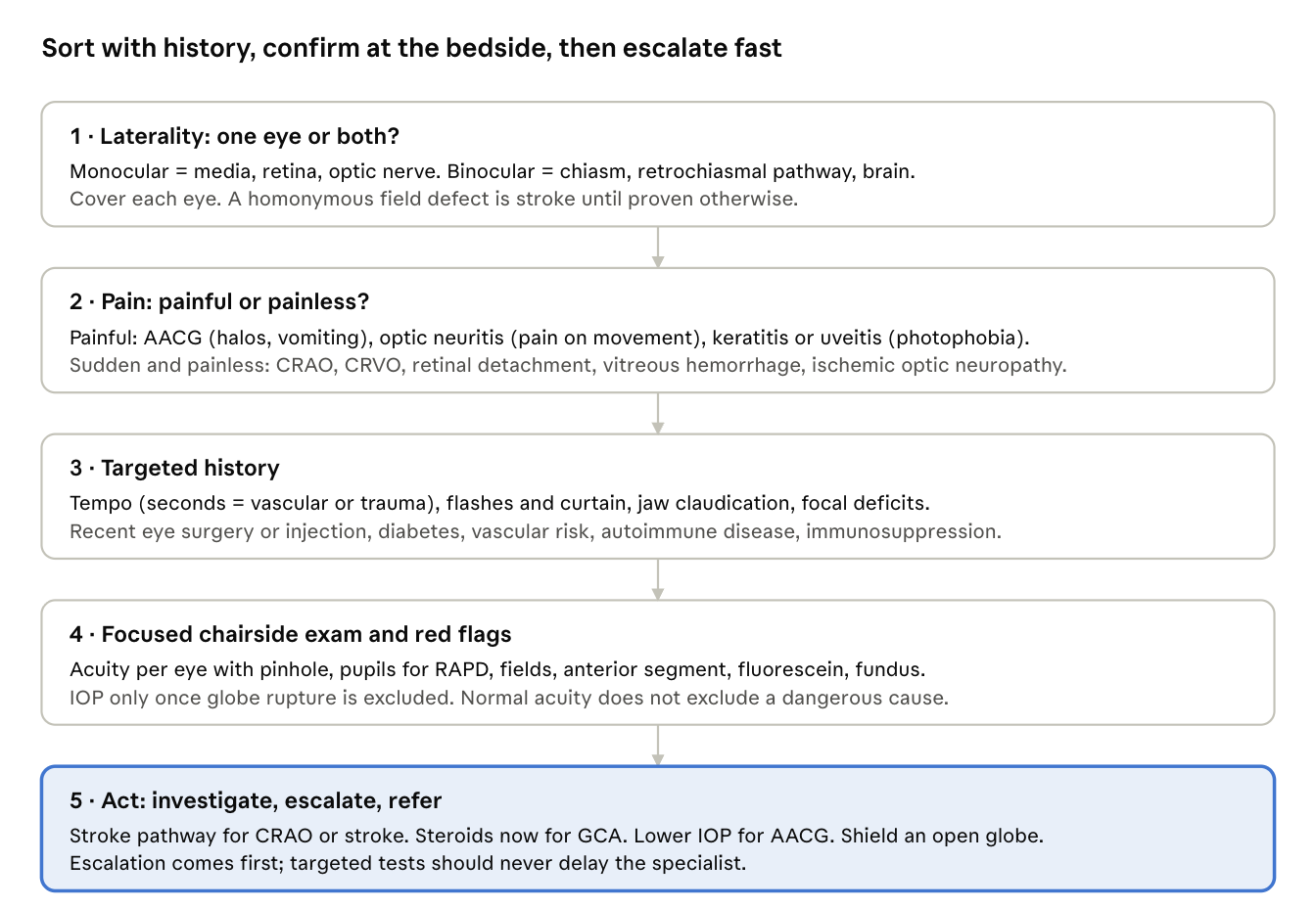
Steps 1–3 narrow the differential in minutes; step 4 confirms red flags at the bedside; step 5 is where outcomes are won or lost.
Summary teaching points
- Use the five steps every time: laterality, pain, targeted history, focused exam with red flags, act.
- Test each eye separately. "Monocular" blur is often a binocular homonymous field defect, which means the brain, not the eye.
- Sudden painless monocular loss is CRAO until proven otherwise. It is a stroke equivalent: activate the stroke pathway, don't just page ophthalmology.
- Over 50 with vision loss and headache or jaw claudication? Think GCA. Draw ESR/CRP, then start steroids without waiting. Normal markers do not rule it out.
- Painful red eye, halos, vomiting, mid-dilated pupil = AACG. Lower the IOP now, call ophthalmology, and never dilate.
- Flashes, floaters and a curtain mean retinal detachment. Preserved central acuity (macula-on) makes it more urgent, not less: same-day ophthalmology.
- Recent eye surgery or injection plus pain and falling vision is endophthalmitis until proven otherwise, especially at 3–5 days.
- Normal acuity does not mean a safe eye. Check pupils for an RAPD and check fields.
- Suspected open globe: shield it, CT the orbits, give antiemetics, keep NPO, update tetanus, and skip tonometry.
- Treat transient loss with the same urgency as persistent loss when the cause could be vascular.
- Don't patch, don't dilate, and don't start topical steroids in the undifferentiated painful red eye.
- Escalation beats diagnosis. The ED goal is to recognize red flags and get the right specialist involved fast; investigations should never delay that.
Reference
Maher C, Guo B, Yek J. A Five-Step Approach to Acute Vision Loss: A Practical Narrative Review for Clinicians. Clinical Ophthalmology. 2026;20:590753. doi:10.2147/OPTH.S590753
This post is an educational summary of a narrative review, not a clinical guideline. Follow local protocols for dosing, thrombolysis eligibility and referral pathways.